


Accurate diagnosis is the first step in treatment.
-
01

At the first visit, we record and analyze a gait video before the consultation.
-
02

A detailed physical exam locates painful or abnormal areas, and possible conditions are ranked by likelihood.
-
03

Radiographs can diagnose more than 90% of conditions.
-
04

When radiographs cannot confirm the diagnosis, MRI, CT or arthroscopy can provide a more accurate one.
- PennHIP & JPS surgery
- Diagnosis with MRI, CT & arthroscopy
PennHIP (Pennsylvania Hip Improvement Program)
A radiographic test that detects hip dysplasia early, so it can be treated: a special device applies force to both thighs, and the looseness (laxity) of the hip joints is measured as a numerical score.
JPS surgery
Juvenile pubic symphysiodesis (JPS) is a surgery that can help prevent hip problems in large-breed puppies identified as high-risk by PennHIP. At the Large Dog Clinic, a PennHIP-certified veterinarian takes and reads the radiographs, supporting an accurate diagnosis.
- For large-breed puppies up to 16 weeks (4 months) of age
- Adjusts the growth plate at the pubic symphysis (where the two halves of the pelvis meet) so that, as the puppy grows, the pelvis covers the femoral head better and hip stability improves
- Helps prevent hip problems in adulthood and may avoid costly surgery such as total hip replacement later on
-

Standard radiograph
No hip problem visible -

PennHIP radiograph
When radiographs are not enough
Pain from a fragmented medial coronoid process (FMCP) in the elbow, partial cruciate ligament tears, meniscal damage or osteochondritis dissecans (OCD) fragments in the shoulder can be missed on radiographs alone; MRI, CT and arthroscopy are more effective for diagnosing them.
At the Large Dog Clinic, arthroscopy is performed by a veterinarian who completed arthroscopy courses in Europe and the US, and experienced veterinarians trained in diagnostic imaging perform and read the MRI and CT scans.
-
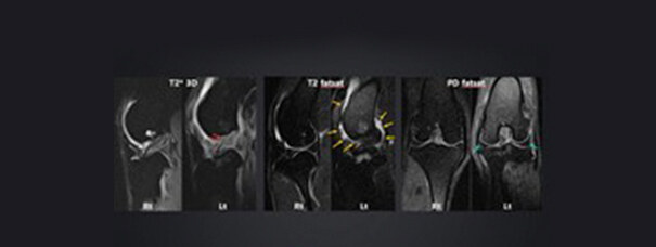
Partial cruciate ligament tear diagnosed on MRI
-

Arthroscopy in progress
Tailored surgical treatment using current techniques
We plan surgery around each patient's condition, drawing on experience with a wide range of surgical cases.-
01Hip dysplasia & degenerative joint disease
Hip disease
Total hip replacement01Hip disease (total hip replacement)
Hip dysplasia & degenerative joint diseaseIn Retrievers, Border Collies, German Shepherds and similar breeds, hip disease accounts for the largest share of hindlimb conditions.
- Total hip replacement (THR) replaces the damaged hip joint entirely with an artificial joint
- Joint replacement aims to restore full joint function
- With femoral head and neck ostectomy (FHNO), a natural gait and complete pain relief can be hard to achieve in large dogs
The Large Dog Clinic has hip replacement implant systems from BioMedtrix (USA) and from Vetmaster and BlueSAO (China). Total hip replacement is performed personally by Dr. Jeong Hee-jun, who holds joint replacement certifications from both the US and China.
Case 01The BioMedtrix Lateral Bolt hip system, and before/after radiographs of a patient with degenerative hip disease
-
 BioMedtrix Lateral Bolt hip replacement system
-
 Before and after radiographs of a patient with degenerative hip disease
-
02Cranial cruciate ligament tear
Cruciate ligament rupture
(TPLO / arthroscopy)02Cruciate ligament rupture (TPLO / arthroscopy)
Cranial cruciate ligament tearWe perform more than 100 TPLO surgeries a year and provide diagnosis, treatment and rehabilitation for cruciate ligament rupture.
- Avoids side effects such as increased patellar ligament tension and limb shortening
- Widely regarded as the gold-standard technique
- When patellar luxation and cruciate rupture occur together, our technique can correct both in a single operation
We use TPLO for cruciate rupture to reduce the risk of long-term gait problems after surgery. Arthroscopy is used to check the meniscus for damage; if it is injured, partial removal or repair of the meniscus helps improve the prognosis.
-
 CTWO surgical plan
-
 TPLO surgical plan
**CTWO (cranial tibial wedge osteotomy) removes a wedge of the tibia, which inevitably shortens the leg and, in rare cases, can leave the hind legs uneven.-
 Before and after TPLO radiographs of a patient with patellar luxation and cruciate rupture at the same time
**Placing pins to correct patellar luxation during TPLO is technically demanding because of the limited bone space. -
03Femoral deformity correction · shoulder and hock arthritis and luxation
Limb deformity
& joint disease03Femoral deformity correction; shoulder & hock arthritis and luxation
(3D-printing-guided angular correction / arthrodesis)Femoral angular deformity correction (3D-printing-guided)
When a congenital deformity makes the femur grow bowed inward, medial patellar luxation and degenerative change in the knee increase the strain on the joint and can lead to hindlimb lameness.
- Surgery is recommended when the angle between the knee joint axis and the long axis of the femur is 105° or more
- The angle is corrected to within the normal range of 95–98°
- We use custom-made CustoMedi 3D-printed guides, licensed as veterinary medical devices
Compared with the old method using a surgical goniometer, 3D-printed guides improve accuracy and stability and shorten operating time.
-
 Making the 3D-printed guide
-
 Radiograph of a femur corrected from 110° before surgery to 95°
Shoulder & hock arthritis and luxation (3D-printing-guided arthrodesis)
Arthritis and luxation of the shoulder or hock can cause severe pain in large dogs. To relieve the pain, the joint surfaces can be removed and the joint fused with a plate (arthrodesis), with 3D printing used to plan and guide the surgery. The goal after surgery is reduced pain and a near-normal gait.
-
 3D-printed guide for shoulder arthrodesis
-
 Radiograph after shoulder arthrodesis
-
04Fracture repair
Fractures
(minimally invasive repair & interlocking nail)04Fractures (minimally invasive repair & interlocking nail)
Fracture repairMinimally invasive fracture surgery, without opening the fracture site or handling the bone fragments, can allow a faster recovery.
The outcome of fracture surgery depends on how closely the bone is restored to its original shape and how little the fracture site is disturbed. The Large Dog Clinic uses minimally invasive plate osteosynthesis (MIPO) and interlocking-nail techniques, which can improve the prognosis.
-
 Before and after radiographs of plate fixation by MIPO, without opening or disturbing the fracture site
-
 Before and after radiographs of interlocking-nail fixation, without opening or disturbing the fracture site
-
-
05Kneecap dislocation
Patellar luxation
05Patellar luxation
Kneecap dislocationLifetime re-luxation policy (free revision surgery) · more than 300 patellar luxation surgeries a year Patellar luxation is less common in large dogs than in small breeds, but large dogs with hip disease or femoral angular deformity are at higher risk. If the kneecap luxates again, the Large Dog Clinic will perform revision surgery free of charge.
-
 (Before and after radiographs of a Retriever with patellar luxation)
The kneecap, previously luxated medially, returned to the center of the femur
-
Inpatient rehabilitation and stem cell therapy to support recovery
Inpatient facilities & tailored rehabilitation
-
 Spacious kennels where large dogs can rest
-
 At least one rooftop walk a day
-
 A large pool, treadmill and other rehabilitation facilities sized for big dogs
Stem cell therapy
-
 Stem cell therapy
-
 Anicon intra-articular injection
Stem cells can differentiate into the same type of cells as the surrounding tissue, so when applied after surgery they can help slow degenerative change in the joint and support the repair of damaged joint tissue. Combined with an Anicon injection, which acts as a cushion inside the joint, they can further support recovery after joint surgery.
Learn more about the Stem Cell Center (SALAB)
