


CT scans
CT (computed tomography) uses X-rays to build three-dimensional images. A short scan yields a great deal of information, and because it is read in 3D — unlike a plain radiograph — lesions anywhere in the body can be examined in fine detail.When a CT scan is needed
Because CT images can be reconstructed in 3D, we use CT when a tumor or vascular disease is suspected. In cancer patients it shows tumor size, invasion and metastasis in detail, and pre-operative CT lets us plan surgery precisely — and, when needed, rehearse it on a 3D-printed model to help improve the chance of success.-

Head & neck
- Otitis externa and media, hydrocephalus, rhinitis, dental disease
- Head and neck tumors, inflammation, hemorrhage
- Sialocele, lymph node enlargement, thyroid assessment
-

Thorax
- Lung, mediastinal, cardiac and esophageal tumors; lung metastasis
- Lung collapse, lung lobe torsion, lymph node enlargement
- Tracheal and bronchial collapse, bronchiectasis
-

Abdomen
- Abdominal organ assessment; abdominal tumors and inflammation
- Hernias; obstruction from gallstones or urinary stones
- Ovarian remnant, retained testicle, congenital ectopic ureter
-

Musculoskeletal & skin
- Skull fractures, occipital dysplasia
- Spinal and rib fractures, disc herniation
- Musculoskeletal and subcutaneous tumors, infiltrative lipoma, chest and abdominal wall defects
-

Vascular disease
- Thrombosis and embolism; congenital and acquired portosystemic shunts
- Patent ductus arteriosus, persistent right aortic arch
Ultrasound
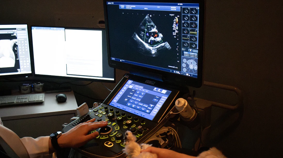
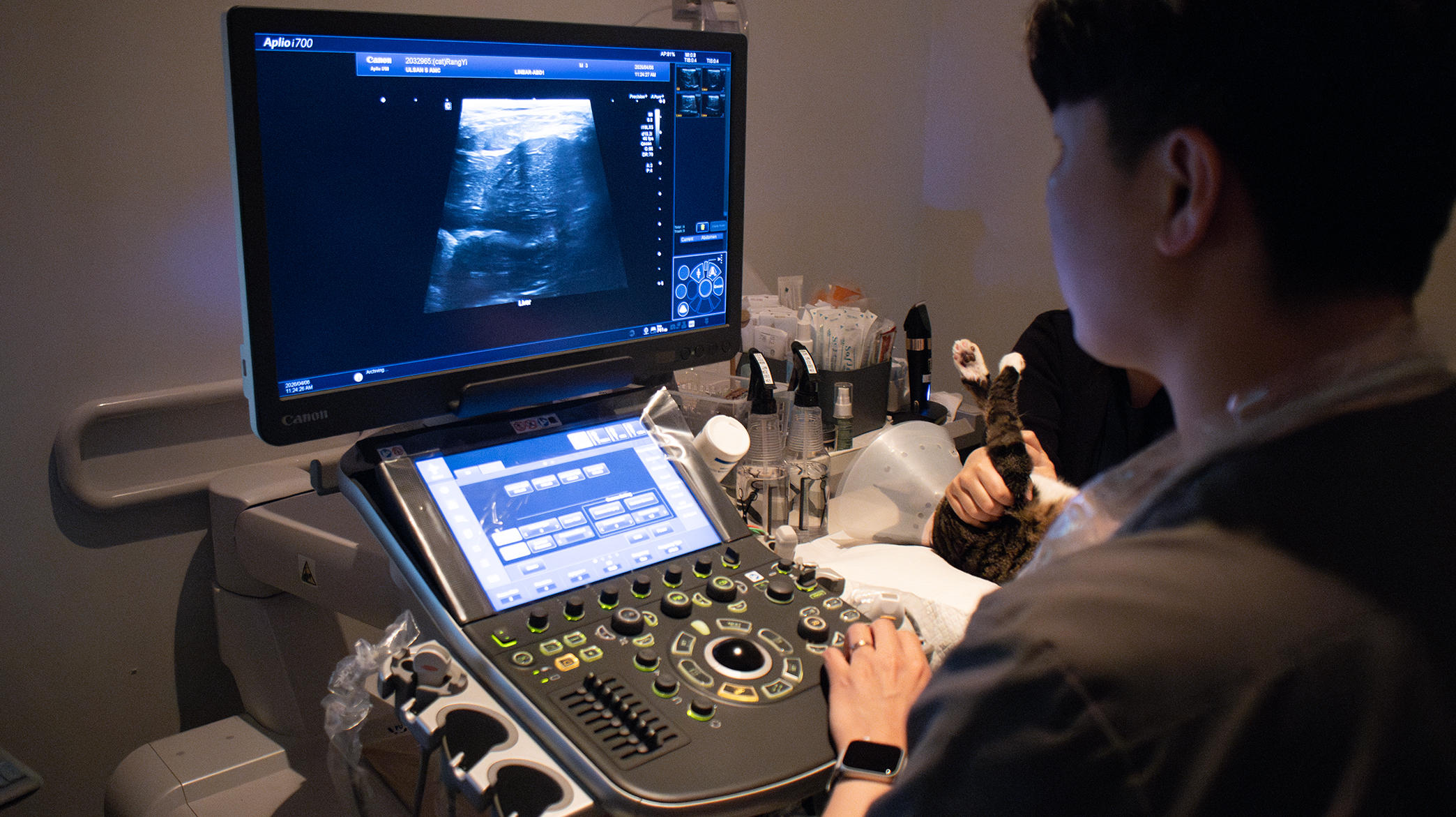
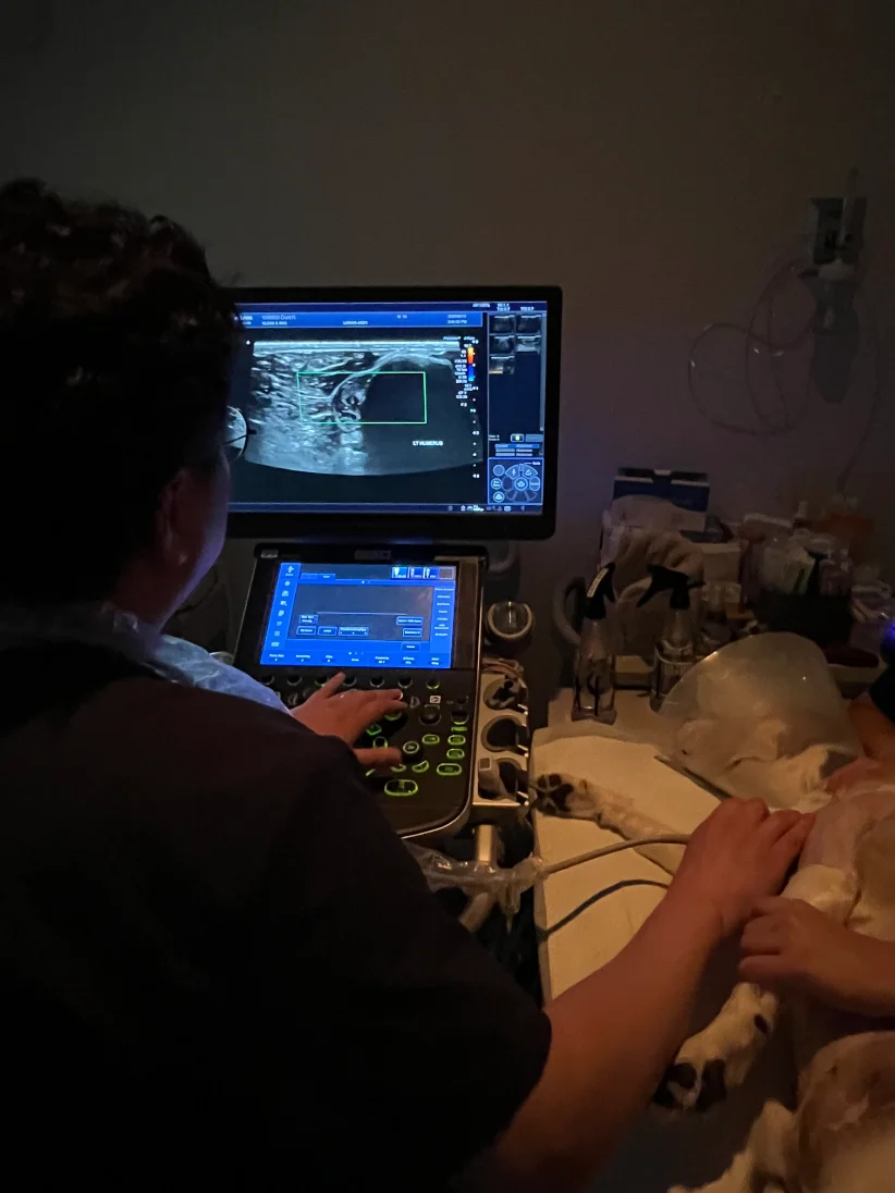
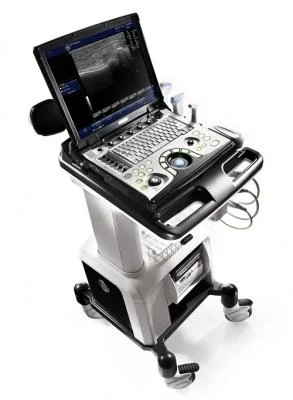
Our ultrasound equipment
Cardiac ultrasound
Assesses the structure and function of the heart and the great vessels of the chest. A key test for diagnosing and monitoring valve disease, contractility, regurgitation and congenital or acquired heart disease; our ultrasound systems can measure heart wall motion for a more detailed evaluation.
- Structure and blood flow: cardiac anatomy and blood flow viewed in real time
Abdominal ultrasound
Evaluates the shape, function and blood flow of the abdominal organs — liver and biliary tract, urinary tract, gastrointestinal and reproductive systems. Also guides procedures such as abdominocentesis and fine-needle aspiration (FNA).
- Detailed organ assessment: liver, spleen, pancreas, kidneys and more
- Tumor and microvascular assessment (SMI): captures fine blood flow for precise evaluation
Musculoskeletal ultrasound
Real-time assessment of joints, ligaments, muscles and tendons.
- Fine-structure diagnosis: the cruciate ligament and tissues around the kneecap
- Soft tissue tumors: maps the blood supply within a tumor and its relationship to surrounding tissue
Ocular ultrasound
When the basic eye examination (intraocular pressure, slit lamp) cannot give an answer, ultrasound shows the structures inside the eye in detail. An ultra-high-frequency hockey-stick probe examines the retina, lens and vitreous closely.
- Retinal detachment and retinal tumors
- Lens luxation or opacity, vitreous hemorrhage, intraocular masses and more

